Virginia Patent of the Month – October 2023
Retinal imaging has been a crucial tool in diagnosing various eye diseases, but the conventional retinal cameras had limitations in capturing a wide field of view. For conditions like retinopathy of prematurity (ROP), which primarily affect the retinal periphery, a minimum field of view of 120 degrees is required for accurate diagnosis. In the past, physicians relied on indirect ophthalmoscopy, which was both time-consuming and lacked reliable documentation.
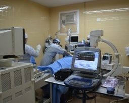
RetiVue, LLC, retinal photography experts, have recently patented their design for a wide field fundus camera. These advanced cameras utilize a range of technologies to provide comprehensive retinal images including an aspherical lens with a symmetric viewing axis. This lens forms a wide field of view retinal image of the subject’s retina.
Positioned around the viewing axis, multiple illumination beam projectors project illumination beams with predetermined profiles and angles. This technique allows precise and even illumination of different sections of the retina within the wide field of view.
Placed between the subject’s eye and the image recording device, a cross polarization system efficiently rejects specular reflections of the illumination beams. This ensures clear and artifact-free images.The camera is equipped with an image recording device capable of auto focus. This feature is particularly valuable for handheld devices, ensuring quick and reliable focusing even in challenging scenarios. Using a processor circuit, the camera stitches together multiple sectional images into a single montage, providing a wide field of view retinal image.
To optimize image clarity through less-transparent crystalline lenses, the camera uses a narrow slit beam projected at an angle. This minimizes reflection haze and enhances auto focus, even in challenging cases.
RetiVue’s wide field fundus camera represents a groundbreaking development in retinal imaging technology. Its ability to provide a comprehensive view of the retina makes it a valuable tool for diagnosing various eye diseases, including those that predominantly affect the retinal periphery like ROP. With this advancement, the field of ophthalmology gains a powerful tool for early disease detection and patient care.
Are you developing new technology for an existing application? Did you know your development work could be eligible for the R&D Tax Credit and you can receive up to 14% back on your expenses? Even if your development isn’t successful your work may still qualify for R&D credits (i.e. you don’t need to have a patent to qualify). To find out more, please contact a Swanson Reed R&D Specialist today or check out our free online eligibility test.
Who We Are:
Swanson Reed is one of the U.S.’ largest Specialist R&D tax advisory firms. We manage all facets of the R&D tax credit program, from claim preparation and audit compliance to claim disputes.
Swanson Reed regularly hosts free webinars and provides free IRS CE and CPE credits for CPAs. For more information please visit us at www.swansonreed.com/webinars or contact your usual Swanson Reed representative.